Educational content only. Not personalised medical advice. Clinical decisions remain the responsibility of the treating clinician.
Optic Neuritis
Optic neuritis is an inflammatory optic neuropathy and the most common optic neuropathy affecting young adults [S1] [S6]. It may be the first sign of multiple sclerosis (MS), neuromyelitis optica spectrum disorder (NMOSD) or myelin oligodendrocyte glycoprotein antibody-associated disease (MOGAD), and these differ in prognosis and treatment [S6] [S7]. Cumulatively, optic neuritis is most often caused by conditions other than MS, and its causes and management vary with geography, treatment availability and ethnic background [S7].
Key points - Separate typical (idiopathic or MS-associated) from atypical optic neuritis early: the cause determines prognosis and treatment [S6] [S8] [S10]. - In typical optic neuritis, high-dose corticosteroids shorten the period of visual loss but do not substantially change the final visual outcome [S1] [S9]. - Optic neuritis with AQP4 antibodies (NMOSD) has a more guarded visual prognosis; prompt plasmapheresis added to intravenous methylprednisolone is recommended, and earlier treatment is associated with better recovery [S9] [S10]. - Test for AQP4-IgG and MOG-IgG in all patients with atypical optic neuritis [S3]. - Infectious causes such as syphilis need early, specific therapy [S10].
Evidence last checked: 2026-10-17 (PubMed abstracts).
Red flags
- Features of atypical optic neuritis, which suggest a less favourable outcome and need special consideration, such as early additional treatment or a very slow steroid taper [S8].
- Poor or absent recovery: recovery of acuity can be poorer, and optic atrophy more severe, with MOG, AQP4 or CRMP5 antibodies than in typical optic neuritis [S6].
- Longitudinally extensive optic nerve inflammation on MRI, seen in both AQP4-associated and MOG-associated optic neuritis [S10].
- Recurrent attacks, typical of MOGAD [S3] [S10].
- Possible infection, sarcoidosis or other granulomatous inflammation: appropriate therapy differs radically from demyelinating optic neuritis [S10].
Recognise it
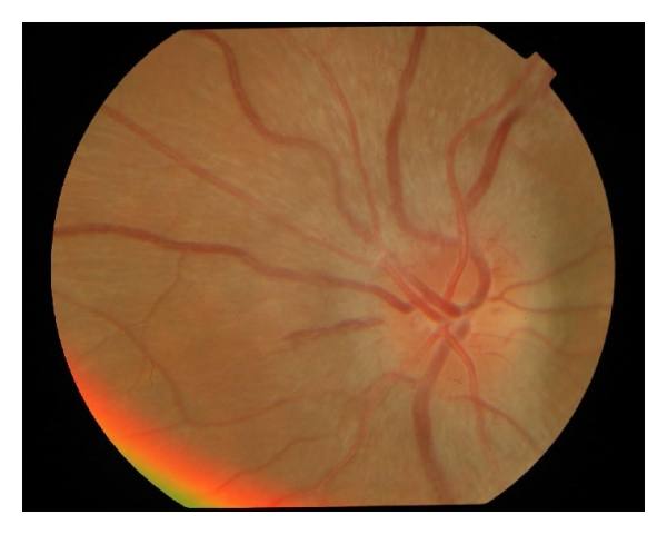
- Typical optic neuritis is an inflammatory demyelinating disorder of the optic nerve that can be associated with MS [S1]. MS-related optic neuritis is the most common form of inflammatory demyelinating optic neuritis and causes short segments of optic nerve inflammation [S10].
- MOGAD optic neuritis has a predilection for optic disc oedema and perineural optic nerve enhancement on MRI, and tends to recur [S3] [S10].
- Atypical optic neuritis includes NMOSD, MOGAD, idiopathic recurrent neuroretinitis, chronic relapsing inflammatory optic neuropathy (CRION), and optic neuritis with systemic autoimmune, paraneoplastic, infectious or post-vaccination causes [S10].
- In children, optic neuritis may be a clinically isolated syndrome or part of MS, NMOSD or MOGAD; distinguishing these guides treatment and long-term prognosis [S4].
- The differential diagnosis includes other optic nerve and retinal disorders [S1].
Confirm it
- An international panel (2022, consensus) proposed criteria in which clinical features give a diagnosis of possible optic neuritis, and brain, orbital and retinal imaging with antibody and other biomarker results give definite optic neuritis [S7].
- Investigations include MRI, visual evoked potentials and CSF examination [S1].
- Antibodies: AQP4-IgG is both the cause and a reliable biomarker of NMOSD; MOG-IgG marks MOGAD [S3].
- Optical coherence tomography (OCT): a meta-analysis of 40 studies (5776 eyes of people with MS) found peripapillary retinal nerve fibre layer thinning of -20.10 µm in eyes after MS-associated optic neuritis and -7.41 µm in MS eyes without optic neuritis, compared with controls; macular ganglion cell and inner plexiform layer thinning was -16.42 µm and -6.31 µm [S2]. The authors recommend these two measures for diagnosis and monitoring [S2]. OCT shows retinal axonal loss that correlates with persistent visual dysfunction [S1].
Manage it
Before acting on this section, check the national and local guidelines, product information and regulatory approvals that apply where you practise. See the medical disclaimer.
Tier A (primary care / no specialist)
- Refer any new unexplained visual loss with suspected optic neuritis to an eye service for diagnosis; the cause decides treatment [S6] [S10].
- Ask about neurological symptoms, infection and systemic autoimmune disease, which point to atypical causes [S10].
Tier B (general eye clinic)
- Record acuity, colour vision, contrast and fields: residual deficits in contrast sensitivity, binocular vision and motion perception can impair quality of life even after acuity recovers [S6].
- Decide whether the picture is typical or atypical [S8].
- Typical optic neuritis: high-dose steroids remain the mainstay of acute treatment [S8]. Intravenous methylprednisolone hastens recovery but does not substantially affect the final outcome [S9]; recovery tends to occur with or without steroids and the visual prognosis is excellent [S10]. Steroid dosing is not given in these abstracts: follow local protocols.
- Atypical optic neuritis: arrange antibody testing and urgent specialist care [S3] [S8].
- Where antibody testing or MRI is not available, discuss with a specialist centre; management varies with treatment availability [S7].
Tier C (specialist centre)
- NMOSD-associated optic neuritis: intravenous methylprednisolone with adjunctive plasmapheresis; shorter time to treatment is associated with higher likelihood of recovery [S9]. Untreated patients have poor visual outcomes [S10].
- MOGAD-associated optic neuritis: natural history untreated is unclear; intravenous methylprednisolone is near-universal and the disease tends to be very steroid sensitive [S9] [S10].
- Other acute options are plasma exchange and intravenous immunoglobulin [S8].
- Long-term treatment: warranted in NMOSD and in MOGAD with poor visual recovery or recurrent attacks [S9]. Most MS disease-modifying agents are ineffective for AQP4-positive NMOSD and MOGAD [S3]. Newer immunosuppressants and monoclonal antibodies have reduced relapses [S5].
- Early, accurate subtyping with timely high-dose corticosteroids and, in some cases, plasmapheresis may preserve acuity, contrast, colour vision and fields [S6].
Refer when
| Situation | Urgency |
|---|---|
| Suspected optic neuritis with atypical features, severe loss or no recovery | Urgent specialist referral [S8] [S9] [EIC expert opinion] |
| Known or suspected NMOSD with new visual loss | Immediate: earlier treatment is associated with better recovery [S9] |
| Bilateral or recurrent optic neuritis, or optic neuritis in a child | Urgent; timing per your local service or specialist advice [S4] [S10] |
| Typical optic neuritis in a young adult | Urgent, for MRI and neurology assessment of MS risk; timing per your local service or specialist advice [S1] [S3] |
Pitfalls
- Treating all optic neuritis as MS-related; worldwide, most is not [S7].
- Starting MS disease-modifying drugs in AQP4-positive NMOSD or MOGAD, where most are ineffective [S3].
- Delaying plasmapheresis in NMOSD-associated optic neuritis [S9].
- Missing syphilis or granulomatous disease [S10].
- Judging recovery by acuity alone [S6].
Evidence notes
| Source | Date | Design |
|---|---|---|
| [S1] | 2014 | Narrative review |
| [S2] | 2017 | Systematic review and meta-analysis, 40 studies; no funding |
| [S3] | 2020 | Narrative review |
| [S4] | 2020 | Narrative review, paediatric |
| [S5] | 2021 | Narrative review (India) |
| [S6] | 2023 | Narrative review, international authors |
| [S7] | 2022 | International consensus diagnostic criteria; require validation |
| [S8] | 2023 | Narrative review |
| [S9] | 2024 | Narrative review |
| [S10] | 2025 | Narrative review with case reports |
- The search sheet held no primary RCT abstract; statements on steroid effect in typical optic neuritis come from reviews [S1] [S9]. The Optic Neuritis Treatment Trial (completed 1991) is mentioned only for MS risk stratification [S3].
- The 2022 diagnostic criteria are consensus-based and need validation [S7].
- Most evidence comes from high-income settings, although causes and management vary by region [S7].
- Not covered by these sources: clinical symptoms such as pain, steroid doses, oral versus intravenous steroids, MS risk figures, and management where MRI or antibody testing is unavailable.
Related articles
References
See the numbered source list below. Each marker [S#] links to its PubMed and DOI record.
Sources
- [S1] Toosy AT, Mason DF, Miller DH Optic neuritis. The Lancet. Neurology. (2014). PubMed 24331795 · doi:10.1016/s1474-4422(13)70259-x
- [S2] Petzold A, Balcer LJ, Calabresi PA, Costello F, Frohman TC, Frohman EM, et al. Retinal layer segmentation in multiple sclerosis: a systematic review and meta-analysis. The Lancet. Neurology. (2017). PubMed 28920886 · doi:10.1016/s1474-4422(17)30278-8
- [S3] Chen JJ, Pittock SJ, Flanagan EP, Lennon VA, Bhatti MT Optic neuritis in the era of biomarkers. Survey of ophthalmology. (2020). PubMed 31425702 · doi:10.1016/j.survophthal.2019.08.001
- [S4] Gise RA, Heidary G Update on Pediatric Optic Neuritis. Current neurology and neuroscience reports. (2020). PubMed 32124097 · doi:10.1007/s11910-020-1024-x
- [S5] Phuljhele S, Kedar S, Saxena R Approach to optic neuritis: An update. Indian journal of ophthalmology. (2021). PubMed 34427197 · doi:10.4103/ijo.ijo_3415_20
- [S6] Bennett JL, Costello F, Chen JJ, Petzold A, Biousse V, Newman NJ, et al. Optic neuritis and autoimmune optic neuropathies: advances in diagnosis and treatment. The Lancet. Neurology. (2023). PubMed 36155661 · doi:10.1016/s1474-4422(22)00187-9
- [S7] Petzold A, Fraser CL, Abegg M, Alroughani R, Alshowaeir D, Alvarenga R, et al. Diagnosis and classification of optic neuritis. The Lancet. Neurology. (2022). PubMed 36179757 · doi:10.1016/s1474-4422(22)00200-9
- [S8] Keyhanian K, Chwalisz BK The Treatment of Acute Optic Neuritis. Seminars in ophthalmology. (2023). PubMed 37162276 · doi:10.1080/08820538.2023.2211662
- [S9] Bergeron E, Bouffard MA Evidence-based management of optic neuritis. Current opinion in ophthalmology. (2024). PubMed 37846574 · doi:10.1097/icu.0000000000001007
- [S10] Eggenberger E Optic Neuritis. Continuum (Minneapolis, Minn.). (2025). PubMed 40179402 · doi:10.1212/cont.0000000000001560