Educational content only. Not personalised medical advice. Clinical decisions remain the responsibility of the treating clinician.
Blepharoptosis
Blepharoptosis (ptosis) is drooping of the upper eyelid; it is among the most common upper eyelid disorders and can affect appearance and visual function [S9] [S2]. Ptosis is a non-specific sign that sometimes heralds a life-threatening neurological disorder [S2], and in children it can cause amblyopia [S7] [S6].
Key points - Measure palpebral fissure height, marginal reflex distance, upper lid crease and levator function [S2]. - In acquired ptosis, check onset, symptoms, pupil size and extraocular muscle function to rule out serious causes; if signs of a serious cause are present, immediate referral or testing is required [S9]. - In congenital ptosis, amblyopia risk, ptosis grade and levator function guide timing and technique [S3]. - For poor levator function, frontalis suspension, frontalis flap and maximal levator resection have overlapping success and exposure rates; evidence is mostly level III [S11].
Evidence last checked: 2026-10-17 (PubMed abstracts).
Red flags
- Pupil involvement, other cranial nerve or extraocular muscle involvement: these features narrow the differential and can point to serious neurological disease [S2] [S9].
- Variable ptosis with diplopia: consider ocular myasthenia gravis, which can cause disabling diplopia or functional blindness from ptosis [S5].
- A child whose lid covers the visual axis: risk of amblyopia, astigmatism and torticollis [S7] [S3].
- Progressive bilateral ptosis with poor eye movements: myogenic causes (CPEO, OPMD) carry loss of corneal protection [S10].
Recognise it
- Categories: pseudoptosis, congenital and acquired; acquired causes are mechanical, myogenic, neuromuscular, neurogenic and cerebral [S2]. Acquired ptosis is the predominant form [S9].
- Congenital ptosis may be unilateral or bilateral and associated with Marcus Gunn, Horner and Duane syndromes [S3]; it often results from poor development of the levator muscle [S7].
- After eye surgery: ptosis is common after ocular surgery, with proposed mechanisms including speculum tension, prostaglandin analogue use and trauma at the tarsal plate [S1].
- After botulinum toxin injection: onset is typically 3 to 14 days after injection and it resolves as the toxin wears off [S8].
Confirm it
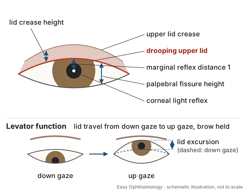
- Measure the four parameters: palpebral fissure height, marginal reflex distance, upper lid crease and levator function [S2].
- Examine pupils, extraocular movements, other cranial nerves and laterality [S2] [S9].
- Exclude masquerade conditions and pseudoptosis before treating [S9].
- In children, assess amblyopia risk [S3] [S6].
- Investigation for myasthenia, Horner syndrome or neuroimaging pathways are not detailed in these abstracts: follow local protocols.
Manage it
Before acting on this section, check the national and local guidelines, product information and regulatory approvals that apply where you practise. See the medical disclaimer.
Tier A (primary care / no specialist)
- Identify ptosis using history, old photographs and lid position and symmetry [S9].
- Check pupils and eye movements; refer immediately if there are signs of a serious underlying cause [S9].
Tier B (general eye clinic)
- Full lid measurements and levator function [S2].
- Children: management needs a structured approach to avoid amblyopia; underlying systemic problems must be identified [S6]. Not every child needs surgery; children at risk of amblyopia need prompt correction, while surgery can be postponed in others [S3].
- Botulinum toxin ptosis: oxymetazoline or apraclonidine eye drops, anticholinesterase agents or pre-tarsal toxin injection can at least partially reverse it [S8].
- Ocular myasthenia: options include pyridostigmine, immunosuppression, lid crutches and ptosis surgery; data comparing treatments are lacking [S5].
- Mild to moderate acquired ptosis: pharmacological or surgical treatment selected on clinical evidence, after excluding serious causes [S9].
Tier C (specialist centre)
- Technique follows levator function. Moderate to good function: external levator advancement or Müllerectomy/Fasanella-Servat; poor function: frontalis sling [S7].
- Severe congenital ptosis (levator function 4 mm or less): an AAO review of 11 studies found overlapping success, exposure and revision rates for frontalis flap, frontalis suspension and maximal levator resection; frontalis suspension had sling infections, extrusions and granulomas [S11].
- Sling material: a Cochrane review of 3 small RCTs (160 participants) could not identify the optimal material [S7]. Autogenous fascia lata is considered the material of choice with low recurrence rates, but needs a second surgical site [S4].
- Myogenic ptosis (CPEO, OPMD, myasthenia): success rates in retrospective series ranged from 60.5% to 100%; the goal is to lift just enough to clear the visual axis while protecting the cornea [S10].
- Before any ocular surgery, include postoperative ptosis in consent [S1].
Refer when
| Situation | Urgency |
|---|---|
| Ptosis with abnormal pupil, diplopia or other cranial nerve signs | Immediate, same-day neurological assessment [S9] |
| Variable ptosis or diplopia suggesting myasthenia | Emergency referral [EIC expert opinion] |
| Child with ptosis covering the pupil (amblyopia risk) | Urgent to paediatric ophthalmology or oculoplastics [S3]; timing per your local service or specialist advice |
| Stable acquired ptosis, normal pupils and motility | Routine, for elective assessment; timing per your local service or specialist advice |
Pitfalls
- Missing a neurological cause by not checking pupils and motility [S2] [S9].
- Treating pseudoptosis or a masquerade condition as ptosis [S9].
- Overcorrecting myogenic ptosis and exposing the cornea [S10].
- Delaying assessment in a child at risk of amblyopia [S3].
Evidence notes
| Source | Date | Design |
|---|---|---|
| [S1] | 2016 | Narrative review |
| [S2] | 2016 | Narrative review (neurology) |
| [S3] | 2017 | Narrative review |
| [S4] | 2018 | Narrative review |
| [S5] | 2018 | Narrative review, expert framework |
| [S6] | 2018 | Narrative review |
| [S7] | 2019 | Cochrane systematic review, 3 RCTs |
| [S8] | 2021 | Narrative review with anatomical dissection |
| [S9] | 2022 | Expert consensus review (optometry) |
| [S10] | 2023 | Literature review of retrospective series |
| [S11] | 2026 | AAO evidence report |
- Thin evidence: apart from one Cochrane review of small trials [S7], the sources are reviews and expert opinion; the AAO report rated 9 of 11 studies level III [S11].
- The optometry consensus [S9] promotes a shift from "detect and refer" to "diagnose and manage", supported by evolving pharmacological options; it is expert opinion, not a systematic review.
- Not covered by these sources: prevalence data, specific drug names and doses for pharmacological ptosis treatment, myasthenia testing, and outcomes in low-resource settings.
Related articles
References
See the numbered source list below. Each marker [S#] links to its PubMed and DOI record.
Sources
- [S1] Godfrey KJ, Korn BS, Kikkawa DO Blepharoptosis following ocular surgery: identifying risk factors. Current opinion in ophthalmology. (2016). PubMed 26632916 · doi:10.1097/icu.0000000000000218
- [S2] Yadegari S Approach to a patient with blepharoptosis. Neurological sciences : official journal of the Italian Neurological Society and of the Italian Society of Clinical Neurophysiology. (2016). PubMed 27329276 · doi:10.1007/s10072-016-2633-7
- [S3] Marenco M, Macchi I, Macchi I, Galassi E, Massaro-Giordano M, Lambiase A Clinical presentation and management of congenital ptosis. Clinical ophthalmology (Auckland, N.Z.). (2017). PubMed 28280295 · doi:10.2147/opth.s111118
- [S4] Lee JH, Kim YD Surgical treatment of unilateral severe simple congenital ptosis. Taiwan journal of ophthalmology. (2018). PubMed 29675342 · doi:10.4103/tjo.tjo_70_17
- [S5] Cornblath WT Treatment of Ocular Myasthenia Gravis. Asia-Pacific journal of ophthalmology (Philadelphia, Pa.). (2018). PubMed 30044061 · doi:10.22608/apo.2018301
- [S6] Weaver DT Current management of childhood ptosis. Current opinion in ophthalmology. (2018). PubMed 30096088 · doi:10.1097/icu.0000000000000508
- [S7] Rosenberg JB, Andersen J, Barmettler A Types of materials for frontalis sling surgery for congenital ptosis. The Cochrane database of systematic reviews. (2019). PubMed 31013353 · doi:10.1002/14651858.cd012725.pub2
- [S8] Nestor MS, Han H, Gade A, Fischer D, Saban Y, Polselli R Botulinum toxin-induced blepharoptosis: Anatomy, etiology, prevention, and therapeutic options. Journal of cosmetic dermatology. (2021). PubMed 34378298 · doi:10.1111/jocd.14361
- [S9] Nichols KK, Malloy KA, Karpecki PM, Bacharach J, Douglas RS, Foster S, et al. Topical Review: An Update of Diagnostic and Management Algorithms for Acquired Blepharoptosis. Optometry and vision science : official publication of the American Academy of Optometry. (2022). PubMed 35058403 · doi:10.1097/opx.0000000000001868
- [S10] Park RB, Akella SS, Aakalu VK A review of surgical management of progressive myogenic ptosis. Orbit (Amsterdam, Netherlands). (2023). PubMed 36178005 · doi:10.1080/01676830.2022.2122514
- [S11] Dagi Glass LR, Aakalu VK, Grob SR, Liu CY, Vagefi MR, Yoon MK, et al. Severe Congenital Ptosis Repair: A Report by the American Academy of Ophthalmology. Ophthalmology. (2026). PubMed 42059847 · doi:10.1016/j.ophtha.2026.03.014