Educational content only. Not personalised medical advice. Clinical decisions remain the responsibility of the treating clinician.
Central Retinal Artery Occlusion: An Ocular Stroke
Central retinal artery occlusion (CRAO) is a form of acute ischaemic stroke that causes severe visual loss and heralds further cerebrovascular and cardiovascular events [S4]. It is a medical emergency [S4]. No treatment has proven benefit in randomised trials [S6] [S11] [S12], so the priorities are fast triage, excluding giant cell arteritis, and secondary prevention [S4] [S9] [S8].
Key points - Treat CRAO as a stroke: systems of care should prioritise early recognition and emergency triage [S4] [S8]. - Giant cell arteritis (GCA) can cause CRAO; treatment must start urgently when GCA is suspected with eye signs, to prevent blindness [S9] [S1]. - Observational individual participant data suggest better recovery with early thrombolysis [S10] [S7], but two phase 3 RCTs within 4.5 hours found no significant benefit over aspirin [S11] [S12]. - Conservative measures such as eyeball massage, oxygen or pressure lowering have no proven benefit; the evidence is uncertain [S6]. - Every patient needs urgent screening and treatment of vascular risk factors [S4] [S8].
Evidence last checked: 2026-10-17 (PubMed abstracts).
Red flags
- Sudden, severe, persistent loss of vision in one eye, the presentation enrolled in the THEIA trial [S11]. CRAO is a medical emergency [S4].
- Headache, scalp tenderness or jaw claudication, especially in women over fifty: suspect GCA [S9]. GCA is an ophthalmic emergency needing early diagnosis and immediate high-dose steroid to protect both eyes [S1].
- Neurological symptoms with the eye event need neurological evaluation [S2].
- High risk of further vascular events. Patients have very high cardiovascular risk [S8] and CRAO is a harbinger of stroke and cardiovascular events [S4].
Recognise it
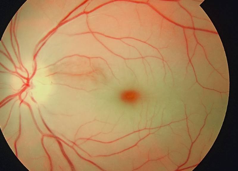
- Retinal artery occlusion is associated with increased age and cardiovascular risk factors [S3].
- Embolism is the most common cause [S2].
- Subtypes differ in prognosis. In eyes seen within 7 days with counting fingers or worse, vision improved spontaneously in 82% of transient non-arteritic CRAO, 67% with cilioretinal artery sparing and 22% of non-arteritic CRAO [S1]. Final acuity of 20/40 or better was seen in 89% of permanent branch retinal artery occlusion [S1].
Confirm it
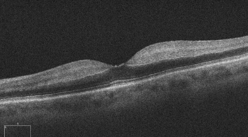
- Record the time of onset: time to treatment influenced visual outcome in observational data [S10] [S7].
- Exclude GCA. Temporal artery biopsy is the gold standard; Doppler ultrasound and vascular imaging are also reliable [S9]. Do not delay treatment for tests when GCA is strongly suspected [S9].
- Find the embolic source. Evaluate the carotid artery and heart, with fasting lipids and a complete blood count [S2].
Manage it
Before acting on this section, check the national and local guidelines, product information and regulatory approvals that apply where you practise. See the medical disclaimer.
Tier A (primary care / no specialist)
- Treat as an emergency: arrange immediate transfer to emergency care, ideally where stroke expertise is available [S4] [S3].
- Ask about GCA symptoms [S9]. If GCA is strongly suspected with eye signs, start treatment urgently; the French protocol reports prednisone 40-80 mg per day as initial treatment (as reported in [S9]). Follow local protocols for steroid route and dose.
- Do not delay transfer for eyeball massage or other conservative measures; none has proven benefit [S6].
- Start risk-factor control with primary care afterwards [S3] [S4].
Tier B (general eye clinic)
- Confirm the diagnosis and subtype, including cilioretinal sparing and branch occlusion, which carry a better prognosis [S1].
- Screen for GCA and arrange urgent treatment [S9].
- Arrange carotid and cardiac evaluation for embolism [S2] and refer for vascular risk-factor treatment [S4] [S8].
- Coordinate care with neurologists with stroke expertise [S3].
Tier C (specialist centre)
- Intravenous thrombolysis: conflicting evidence. - In an individual participant data meta-analysis of 783 patients, intravenous thrombolysis within 4.5 hours was associated with recovery from severe vision loss in 28.8% vs 11.1% with conservative therapy (odds ratio 3.32); each hour earlier was associated with better acuity [S10]. Another meta-analysis of 771 patients reported 0.3 logMAR or more improvement in 74.3% after intravenous tPA within 4.5 hours [S7]. Both used non-randomised data. - THEIA (France, 70 patients): alteplase 0.9 mg/kg vs aspirin 300 mg within 4.5 hours (as reported in [S11]) gave improvement in 66% vs 48%, not statistically significant; the trial was likely underpowered and had no symptomatic haemorrhages [S11]. - TenCRAOS (78 patients, six countries): tenecteplase 0.25 mg/kg vs aspirin 300 mg (as reported in [S12]) gave vision recovery in 20% vs 24% at 30 days, with more adverse events including one fatal intracranial haemorrhage [S12]. - The AHA statement (2021, expert consensus) concluded that intravenous tPA may be effective [S4]. Benefit has not been confirmed in RCTs [S11] [S12].
- Intra-arterial thrombolysis. A meta-analysis of 5 studies found any visual improvement more likely within 6 hours of onset (odds ratio 1.703) but not beyond [S5]. Individual participant data showed a changepoint at 8 hours [S10].
- Other interventions (hemodilution, pentoxifylline, transcorneal electrical stimulation) showed no clear benefit in small RCTs [S6].
Refer when
| Situation | Urgency |
|---|---|
| Sudden severe painless loss of vision in one eye | Immediate transfer to emergency care with stroke expertise [S4] [S3] |
| Visual loss with headache, scalp tenderness or jaw claudication | Immediate, same-day treatment for suspected GCA [S9] |
| Visual loss with neurological symptoms | Immediate neurological and stroke assessment [S2] |
| Confirmed retinal artery occlusion, any type | Urgent vascular work-up; timing per your local service or specialist advice [S4] [S8] |
Pitfalls
- Treating CRAO as an eye-only problem and missing the stroke risk [S4].
- Missing GCA, which threatens the fellow eye [S1] [S9].
- Overstating thrombolysis: observational data are positive but RCTs are not [S10] [S11] [S12].
- Wasting time on unproven conservative manoeuvres [S6].
Evidence notes
| Source | Date | Design |
|---|---|---|
| [S1] | 2014 | Prospective natural history studies, single investigator |
| [S2] | 2018 | Literature review and perspective |
| [S3] | 2020 | Narrative review |
| [S4] | 2021 | AHA scientific statement, expert consensus |
| [S5] | 2022 | Meta-analysis, 5 studies |
| [S6] | 2023 | Cochrane systematic review, 6 small RCTs |
| [S7] | 2024 | Individual participant data meta-analysis, 72 publications |
| [S8] | 2024 | Consensus position paper (Spain) |
| [S9] | 2025 | National GCA protocol (France) |
| [S10] | 2025 | Individual participant data meta-analysis, 783 patients |
| [S11] | 2025 | Phase 3 RCT (THEIA), 70 patients; part industry-funded |
| [S12] | 2026 | Phase 3 RCT (TenCRAOS), 78 patients |
- Conflict: observational meta-analyses favour early thrombolysis [S7] [S10]; the two RCTs did not show benefit and one raised safety concerns [S11] [S12]. A trial would need about 95 participants per group to detect an odds ratio of 3.0 [S10].
- Conflict: one expert review recommends carotid and cardiac evaluation rather than neurological evaluation unless there are neurological symptoms [S2]; other sources favour a multidisciplinary stroke approach [S3] [S4].
- Spontaneous improvement occurs in all subtypes, which complicates uncontrolled studies [S1].
- Not covered by these sources: antiplatelet choice and duration, hyperbaric oxygen, neovascular complications after CRAO, and options where no stroke unit is available.
Related articles
References
See the numbered source list below. Each marker [S#] links to its PubMed and DOI record.
Sources
- [S1] Hayreh SS Ocular vascular occlusive disorders: natural history of visual outcome. Progress in retinal and eye research. (2014). PubMed 24769221 · doi:10.1016/j.preteyeres.2014.04.001
- [S2] Hayreh SS Do Patients With Retinal Artery Occlusion Need Urgent Neurologic Evaluation? American journal of ophthalmology. (2018). PubMed 30114396 · doi:10.1016/j.ajo.2018.08.015
- [S3] Scott IU, Campochiaro PA, Newman NJ, Biousse V Retinal vascular occlusions. Lancet (London, England). (2020). PubMed 33308475 · doi:10.1016/s0140-6736(20)31559-2
- [S4] Mac Grory B, Schrag M, Biousse V, Furie KL, Gerhard-Herman M, Lavin PJ, et al. Management of Central Retinal Artery Occlusion: A Scientific Statement From the American Heart Association. Stroke. (2021). PubMed 33677974 · doi:10.1161/str.0000000000000366
- [S5] Hu H, Zhang B, Zhao Y, Zhou H, Chen H, Yan S Efficacy of Intra-Arterial Thrombolysis for Acute Central Retinal Artery Occlusion: A Meta-Analysis. European neurology. (2022). PubMed 34847561 · doi:10.1159/000520443
- [S6] Lin JC, Song S, Ng SM, Scott IU, Greenberg PB Interventions for acute non-arteritic central retinal artery occlusion. The Cochrane database of systematic reviews. (2023). PubMed 36715340 · doi:10.1002/14651858.cd001989.pub3
- [S7] Shahjouei S, Bavarsad Shahripour R, Dumitrascu OM Thrombolysis for central retinal artery occlusion: An individual participant-level meta-analysis. International journal of stroke : official journal of the International Stroke Society. (2024). PubMed 37424312 · doi:10.1177/17474930231189352
- [S8] García-Alonso R, Arias-Barquet L, Castilla Guerra L, Martín Asenjo M, Gómez-Escobar AJ, Gutierrez-Sánchez E, et al. Position paper on retinal arterial occlusion. SEMI-SERV. Revista clinica espanola. (2024). PubMed 39236981 · doi:10.1016/j.rceng.2024.09.001
- [S9] de Boysson H, Devauchelle-Pensec V, Agard C, André M, Bienvenu B, Bonnotte B, et al. French protocol for the diagnosis and management of giant cell arteritis. La Revue de medecine interne. (2025). PubMed 39487062 · doi:10.1016/j.revmed.2024.10.011
- [S10] Xie JS, Zaslavsky K, Chaban YV, Lusterio A, Kaur H, Motekalem Y, et al. Early Thrombolysis and Outcomes in Central Retinal Artery Occlusion: An Individual Participant Data Meta-Analysis. Stroke. (2025). PubMed 40832714 · doi:10.1161/strokeaha.124.049955
- [S11] Préterre C, Gaultier A, Obadia M, Vignal C, Mourand I, Plat J, et al. Intravenous alteplase versus oral aspirin for acute central retinal artery occlusion within 4·5 h of severe vision loss (THEIA): a multicentre, double-dummy, patient-blinded and assessor-blinded, randomised, controlled, phase 3 trial. The Lancet. Neurology. (2025). PubMed 41109232 · doi:10.1016/s1474-4422(25)00308-4
- [S12] Ryan SJ, Jørstad ØK, Skjelland M, Pesonen M, Simonsen CZ, Bek T, et al. A Randomized Trial of Tenecteplase in Acute Central Retinal Artery Occlusion. The New England journal of medicine. (2026). PubMed 41604638 · doi:10.1056/nejmoa2508515