Educational content only. Not personalised medical advice. Clinical decisions remain the responsibility of the treating clinician.
Blunt eye injury and traumatic hyphaema
Blunt (closed-globe) trauma can injure the anterior segment, lens, retina and optic nerve. Traumatic hyphaema, the entry of blood into the anterior chamber after significant injury, can cause raised intraocular pressure (IOP), corneal blood staining, synechiae and optic atrophy, although permanent vision loss is uncommon [S7]. Trauma is a leading cause of monocular blindness, and a structured history and examination decide who can be reassured and who needs immediate referral [S6] [S8].
Key points - First decide whether the globe may be open; a suspected open globe needs immediate referral [S6]. - No medical treatment for hyphaema has been shown to improve visual acuity [S7]. - Antifibrinolytics (aminocaproic acid, tranexamic acid) reduce rebleeding, but the evidence is of low certainty and their effect on complications is unclear [S7]. - People with sickle cell trait or disease are especially susceptible to IOP rise and optic atrophy after hyphaema [S7]. - Look for late consequences: traumatic cataract, retinal detachment and cyclodialysis cleft with hypotony [S9] [S11] [S12].
Evidence last checked: 2026-10-17 (PubMed abstracts).
Red flags
- Possible open globe within an apparently blunt injury: needs immediate referral for surgical repair [S6].
- Relative afferent pupillary defect (RAPD) or poor vision: check acuity and the pupils in every injured eye [S2].
- Sight-threatening retrobulbar haemorrhage: lateral canthotomy with cantholysis should be performed immediately [S2].
- Hyphaema in a person with sickle cell trait or disease [S7] [S5].
- Rebleeding, which increases the rate and severity of complications [S7].
- Infant or young child with retinal haemorrhages: multilayered, too numerous to count and extending from the posterior pole to the ora serrata are highly suspicious for abusive head trauma [S10].
Recognise it
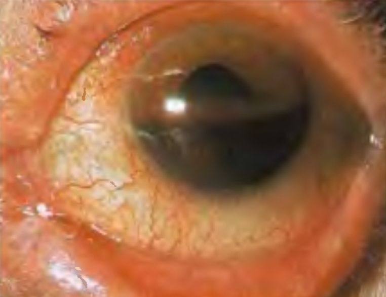
- Common causes include sports, work-related trauma, road traffic accidents and explosions [S3] [S11]. Sports eye injuries have an estimated incidence near 15% [S3].
- Ocular trauma may involve the ocular surface and adnexa, extraocular muscles, orbital walls, eye and optic nerve [S6].
- Hyphaema: blood in the anterior chamber after injury [S7].
- Lens injury: common after blunt or penetrating trauma and can impair vision [S9].
- Cyclodialysis cleft: detachment of the ciliary muscle from the scleral spur, most often after blunt trauma; it may cause persistent severe hypotony with choroidal detachment, folds, disc oedema and macular oedema [S1] [S12].
- Traumatic retinal detachment is a leading cause of trauma-related blindness worldwide [S11].
- Children: abusive head trauma can produce retinal haemorrhages, retinoschisis, subconjunctival haemorrhages, corneal injury and globe rupture [S10].
Confirm it
- Take a detailed history, then a complete ocular examination and, if indicated, imaging [S6].
- Measure visual acuity and test for an RAPD; examine under anaesthesia if necessary [S2].
- Bedside ultrasound can help diagnose vitreous haemorrhage, retinal and vitreous detachment, foreign bodies and retrobulbar haematoma when the fundus cannot be seen [S4].
- Cyclodialysis clefts are hard to diagnose; ultrasound biomicroscopy and anterior segment OCT can help, and more than one cleft may be present [S1].
- Suspected abusive head trauma: an ophthalmology consultation with indirect ophthalmoscopy should be completed, together with full examination, imaging and laboratory tests to exclude accidental and systemic causes [S10].
Manage it
Before acting on this section, check the national and local guidelines, product information and regulatory approvals that apply where you practise. See the medical disclaimer.
Tier A (primary care / no specialist)
- Use a structured approach to history and examination to decide on reassurance or referral [S6].
- Protect a possibly ruptured globe with an eye shield and refer [S2].
- For hyphaema, non-drug measures such as patching, bed rest and head elevation are not supported by evidence: single versus binocular patching and bed rest versus walking made no difference to rebleeding [S7]. Refer any hyphaema to an eye clinic (see table).
Tier B (general eye clinic)
- Hyphaema, medical options (Cochrane review, 30 trials, 2969 participants) [S7]:
- No intervention improved visual acuity in the short or long term (low or very low certainty).
- Systemic tranexamic acid reduced secondary haemorrhage (RR 0.33; 7 trials, 754 participants), and systemic aminocaproic acid did so too (RR 0.28; 6 trials), although the aminocaproic acid result weakened in a sensitivity analysis.
- Aminocaproic acid increased nausea and vomiting and lengthened the time for the hyphaema to clear.
- Evidence on corticosteroids, cycloplegics and aspirin is too limited to support their use.
- Doses are not covered in the abstract; follow local formularies.
- Monitor IOP, rebleeding and corneal staining, with particular care in sickle cell disease [S7]. A narrative review offers a pragmatic approach, including management in children and sickle cell anaemia [S5].
Tier C (specialist centre)
- Traumatic cataract: the approach depends on age, mechanism of trauma and other conditions; early management matters in children because of amblyopia, inflammation and vitreoretinal adhesions [S9].
- Cyclodialysis cleft: a step-wise approach, conservative first, then laser or cryotherapy and surgery (direct cyclopexy, external plombage, vitrectomy with gas); there are too few studies to judge the safety and efficacy of these techniques [S1]. The aim is reattachment of the ciliary body and closure of the cleft [S12].
- Traumatic retinal detachment: managed by vitreoretinal services; there is no standardised definition beyond a preceding history of trauma [S11].
- A poor initial visual acuity does not guarantee a poor final result; the outcome can be predicted after approximately 3 to 4 weeks [S2].
Refer when
| Situation | Urgency |
|---|---|
| Suspected open globe | Immediate [S6] |
| Sight-threatening retrobulbar haemorrhage | Immediate canthotomy and cantholysis on site, then transfer [S2] |
| Any traumatic hyphaema | Same-day eye clinic assessment |
| Hyphaema with sickle cell trait or disease, rebleeding or raised IOP | Immediate, same-day specialist care [S7] |
| Flashes, floaters or a field defect after blunt trauma (possible retinal detachment) | Urgent, within hours |
| Retinal haemorrhages in an infant | Same-day ophthalmology consultation and child-protection pathway [S10] |
Pitfalls
- Missing an open globe hidden within a blunt injury [S6].
- Expecting patching or bed rest to prevent rebleeding [S7].
- Forgetting sickle cell status in hyphaema [S7].
- Judging the final prognosis on the first visual acuity [S2].
- Overlooking abusive head trauma in an infant with retinal haemorrhages [S10].
Evidence notes
| Source | Date | Design |
|---|---|---|
| [S1] | 2010 | Narrative review (cyclodialysis) |
| [S2] | 2011 | Narrative review (eye injury, blast) |
| [S3] | 2013 | Narrative review (sports) |
| [S4] | 2014 | Narrative review (bedside ultrasound) |
| [S5] | 2016 | Narrative review (hyphaema) |
| [S6] | 2022 | Narrative review (primary care) |
| [S7] | 2023 | Cochrane systematic review, 30 trials |
| [S8] | 2023 | Narrative review (emergency department) |
| [S9] | 2024 | Narrative review (traumatic cataract) |
| [S10] | 2024 | Narrative review (abusive head trauma) |
| [S11] | 2025 | Narrative review (traumatic retinal detachment) |
| [S12] | 2025 | Narrative review (cyclodialysis) |
- Thin evidence base. Only the Cochrane review [S7] is a systematic synthesis; all other sources are narrative reviews (expert opinion). Two abstracts give only a scope without findings [S5] [S8].
- The reduction in rebleeding with antifibrinolytics did not translate into a proven benefit for vision, and the long-term effects of secondary haemorrhage are unknown [S7].
- Not covered by these sources: IOP-lowering drug choices in hyphaema and sickle cell disease, surgical washout criteria, commotio retinae, choroidal rupture, angle recession glaucoma and follow-up intervals.
Related articles
References
See the numbered source list below. Each marker [S#] links to its PubMed and DOI record.
Sources
- [S1] Ioannidis AS, Barton K Cyclodialysis cleft: causes and repair. Current opinion in ophthalmology. (2010). PubMed 20051856 · doi:10.1097/icu.0b013e3283366a4d
- [S2] Scott R The injured eye. Philosophical transactions of the Royal Society of London. Series B, Biological sciences. (2011). PubMed 21149360 · doi:10.1098/rstb.2010.0234
- [S3] Aerni GA Blunt visual trauma. Clinics in sports medicine. (2013). PubMed 23522510 · doi:10.1016/j.csm.2012.12.005
- [S4] Kilker BA, Holst JM, Hoffmann B Bedside ocular ultrasound in the emergency department. European journal of emergency medicine : official journal of the European Society for Emergency Medicine. (2014). PubMed 24002686 · doi:10.1097/mej.0000000000000070
- [S5] Bansal S, Gunasekeran DV, Ang B, Lee J, Khandelwal R, Sullivan P, et al. Controversies in the pathophysiology and management of hyphema. Survey of ophthalmology. (2016). PubMed 26632664 · doi:10.1016/j.survophthal.2015.11.005
- [S6] Heath Jeffery RC, Dobes J, Chen FK Eye injuries: Understanding ocular trauma. Australian journal of general practice. (2022). PubMed 35773155 · doi:10.31128/ajgp-03-21-5921
- [S7] Woreta FA, Lindsley KB, Gharaibeh A, Ng SM, Scherer RW, Goldberg MF Medical interventions for traumatic hyphema. The Cochrane database of systematic reviews. (2023). PubMed 36912744 · doi:10.1002/14651858.cd005431.pub5
- [S8] Bae E, Messman A, Shah K Ocular injuries: emergency department strategies. Emergency medicine practice. (2023). PubMed 37768702
- [S9] Soleimani M, Cheraqpour K, Salari F, Fadakar K, Habeel S, Baharnoori SM, et al. All about traumatic cataracts: narrative review. Journal of cataract and refractive surgery. (2024). PubMed 38350230 · doi:10.1097/j.jcrs.0000000000001424
- [S10] Jain D, Le L, Kissoon N Ophthalmologic findings in abusive head trauma. Seminars in pediatric neurology. (2024). PubMed 38964817 · doi:10.1016/j.spen.2024.101141
- [S11] Chauhan K, Dave VP, de Ribot FM, Agrawal R, Sallam AB, Andayani G, et al. Traumatic retinal detachment: A contemporary update. Survey of ophthalmology. (2025). PubMed 39222801 · doi:10.1016/j.survophthal.2024.08.008
- [S12] Sethi A, Udenia H, Beri N, Angmo D, Bari A, Sharma N, et al. Surgical Management of Cyclodialysis Cleft: An Update. Journal of current glaucoma practice. (2025). PubMed 41113793 · doi:10.5005/jp-journals-10078-1469