Educational content only. Not personalised medical advice. Clinical decisions remain the responsibility of the treating clinician.
Rhegmatogenous Retinal Detachment
Rhegmatogenous retinal detachment (RRD) is the most common form of retinal detachment: a retinal break lets fluid pass from the vitreous cavity into the subretinal space, separating the retina [S1] [S4]. It occurs in about 1 in 10,000 people a year [S1], and macula-involving RRD is one of the most common ophthalmic surgical emergencies [S11]. Vitrectomy, scleral buckling and pneumatic retinopexy are all accepted repairs with broadly similar final outcomes [S4] [S6].
Key points - Macula-off detachment carries worse vision and more distortion after repair than macula-on detachment [S7] [S10]. - Point-of-care ultrasound is 94.2% sensitive and 96.3% specific for RRD when the fundus view is limited or no ophthalmologist is available [S3]. - Vitrectomy and scleral buckling give similar reattachment and vision; vitrectomy causes more cataract and new breaks, buckling more choroidal detachment [S4] [S6]. - Pneumatic retinopexy, in selected eyes, gave better vision but lower single-operation success than vitrectomy in the PIVOT RCT [S2]. - No drug to prevent proliferative vitreoretinopathy (PVR) is established [S12].
Evidence last checked: 2026-10-17 (PubMed abstracts).
Red flags
- Detachment that has reached or threatens the macula. Macular detachment was associated with worse postoperative vision [S7], and metamorphopsia was 2.88 times more frequent in macula-off than macula-on RRD [S10].
- Signs of PVR. In children, final success was about 25% lower in eyes with PVR [S8].
- Children and congenital anomalies. Paediatric RRD has delayed diagnosis and more complications; final success was about 36% lower with congenital anomalies [S8].
Recognise it
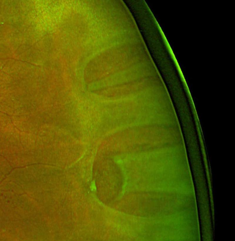
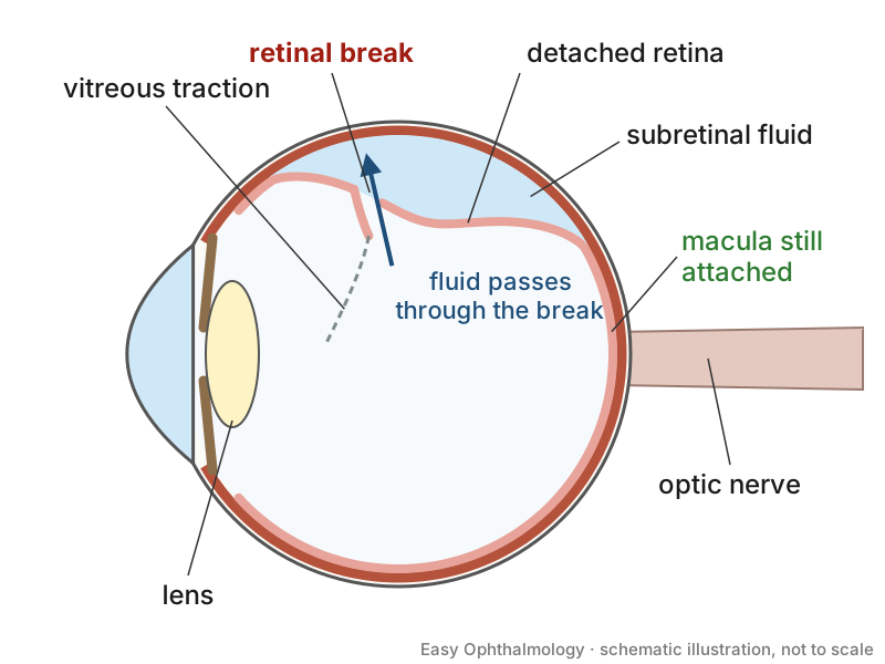
- RRD is caused by a full-thickness retinal break associated with vitreous traction [S5].
- In the OCT meta-analysis, 89% of eyes were macula-off at presentation and mean duration of detachment was 15 ± 10 days [S7].
Confirm it
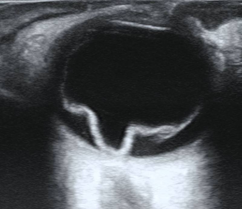
- Ultrasound where the view is poor or no ophthalmologist is available. In 11 studies (844 patients), point-of-care ultrasound had a positive likelihood ratio of 25.2 and a negative likelihood ratio of 0.06; accuracy was higher among emergency department patients [S3].
- Preoperative OCT for prognosis. Greater detachment height at the fovea, disrupted ellipsoid zone or external limiting membrane (vision worse by 0.35 logMAR) and intraretinal cysts were associated with poorer postoperative vision (low-quality evidence) [S7].
Manage it
Before acting on this section, check the national and local guidelines, product information and regulatory approvals that apply where you practise. See the medical disclaimer.
Tier A (primary care / no specialist)
- Suspected RRD is a surgical emergency [S11]: refer to the nearest vitreoretinal service (see Refer when).
- If trained and equipped, point-of-care ultrasound can support the diagnosis; accuracy did not differ significantly between emergency and non-emergency providers [S3].
Tier B (general eye clinic)
- Confirm the diagnosis and macular status; use ultrasound if the view is poor [S3].
- Record whether the macula is on or off, because it affects prognosis and urgency [S7] [S10] [S2].
- Arrange transfer to a vitreoretinal surgeon (see Refer when).
Tier C (specialist centre)
- Vitrectomy vs scleral buckling (simple RRD). A Cochrane review of 10 RCTs (1,307 eyes) found little or no difference in reattachment or vision, fewer redetachments with vitrectomy (risk ratio 0.75), more cataract (risk ratio 1.71) and new breaks with vitrectomy, and more choroidal detachment with buckling (all low certainty) [S4]. A meta-analysis of 15,947 eyes found similar primary (86.5% vs 84.8%) and final reattachment, with a slightly better final acuity after buckling driven by observational studies and phakic eyes [S6].
- Pneumatic retinopexy. In PIVOT (176 patients with a single break or breaks within 1 clock hour in the upper retina), pneumatic retinopexy gave 4.9 letters better vision at 12 months and less metamorphopsia, but lower primary success (80.8% vs 93.2%); secondary success was about 99% in both groups, and 65% of phakic eyes after vitrectomy vs 16% after pneumatic retinopexy underwent cataract surgery within 12 months [S2]. A meta-analysis of 6 comparative studies found higher primary reattachment with vitrectomy and similar final success and vision [S9]. Against buckling, pneumatic retinopexy may reattach slightly less often but with fewer complications (low certainty) [S5].
- Intraoperative choices. Perfluorocarbon liquid during vitrectomy reduced metamorphopsia (risk ratio 0.61) [S10]. Gas vs silicone oil made little or no difference to metamorphopsia [S10].
- Positioning. After vitrectomy with gas for macula-involving RRD, immediate face-down positioning may reduce retinal displacement, outer retinal folds and diplopia but may increase raised intraocular pressure (very low certainty) [S11].
- PVR. In a network meta-analysis of 23 studies, most drugs did not improve reattachment or reduce PVR recurrence; only 13-cis-retinoic acid showed a significant effect, needing confirmation [S12].
- Children. Single-operation success was about 64% and final success about 84% [S8].
Refer when
| Situation | Urgency |
|---|---|
| Suspected or confirmed RRD with the macula still attached | Immediate, same-day transfer to a vitreoretinal surgeon; PIVOT treated macula-on eyes within 24 hours [S2] |
| Macula-off RRD | Emergency, within 24 hours; PIVOT treated macula-off eyes within 72 hours [S2] [EIC expert opinion] |
| Pain, high pressure or vision loss after RRD surgery | Same-day review by the surgical team |
Pitfalls
- Assuming one operation suits every RRD: trade-offs differ by method [S4] [S5] [S9].
- Forgetting cataract after vitrectomy in phakic eyes [S2] [S4].
- Relying on unproven anti-PVR drugs [S12].
- Delayed presentation, especially in children [S7] [S8].
Evidence notes
| Source | Date | Design |
|---|---|---|
| [S1] | 2014 | Systematic review (BMJ Clinical Evidence), 14 studies |
| [S2] | 2019 | RCT (PIVOT), 176 patients |
| [S3] | 2019 | Diagnostic accuracy meta-analysis, 11 studies |
| [S4] | 2019 | Cochrane systematic review, 10 RCTs |
| [S5] | 2021 | Cochrane systematic review, 3 RCTs |
| [S6] | 2022 | Meta-analysis, 8 RCTs and 33 observational studies |
| [S7] | 2023 | Meta-analysis of 29 observational studies |
| [S8] | 2023 | Meta-analysis, paediatric RRD |
| [S9] | 2023 | Meta-analysis of 6 comparative studies |
| [S10] | 2024 | Meta-analysis, 12 studies |
| [S11] | 2024 | Cochrane systematic review, 3 RCTs |
| [S12] | 2025 | Network meta-analysis, 23 studies |
- Thin evidence on recognition and timing: none of the selected abstracts describes presenting symptoms (such as flashes, floaters or a field shadow), the risk after posterior vitreous detachment, prophylaxis of retinal tears, or the optimal timing of surgery; referral timings above are conservative and unsourced.
- Most RRD trials have high or unclear risk of bias and low-certainty evidence [S4] [S5]. PIVOT results apply only to eyes meeting its break criteria [S2].
- The vitrectomy vs buckling meta-analysis mixed RCTs and observational studies, with different results by design [S6].
- Not covered by these sources: laser or cryotherapy for retinal tears, giant retinal tears, tractional and exudative detachment, and cost.
Related articles
- Sudden painless vision loss: triage
- Ocular ultrasound and point-of-care ultrasound
- Complications of cataract surgery
References
See the numbered source list below. Each marker [S#] links to its PubMed and DOI record.
Sources
- [S1] Steel D Retinal detachment. BMJ clinical evidence. (2014). PubMed 24807890
- [S2] Hillier RJ, Felfeli T, Berger AR, Wong DT, Altomare F, Dai D, et al. The Pneumatic Retinopexy versus Vitrectomy for the Management of Primary Rhegmatogenous Retinal Detachment Outcomes Randomized Trial (PIVOT). Ophthalmology. (2019). PubMed 30468761 · doi:10.1016/j.ophtha.2018.11.014
- [S3] Gottlieb M, Holladay D, Peksa GD Point-of-Care Ocular Ultrasound for the Diagnosis of Retinal Detachment: A Systematic Review and Meta-Analysis. Academic emergency medicine : official journal of the Society for Academic Emergency Medicine. (2019). PubMed 30636351 · doi:10.1111/acem.13682
- [S4] Znaor L, Medic A, Binder S, Vucinovic A, Marin Lovric J, Puljak L Pars plana vitrectomy versus scleral buckling for repairing simple rhegmatogenous retinal detachments. The Cochrane database of systematic reviews. (2019). PubMed 30848830 · doi:10.1002/14651858.cd009562.pub2
- [S5] Sena DF, Kilian R, Liu SH, Rizzo S, Virgili G Pneumatic retinopexy versus scleral buckle for repairing simple rhegmatogenous retinal detachments. The Cochrane database of systematic reviews. (2021). PubMed 34762741 · doi:10.1002/14651858.cd008350.pub3
- [S6] Dhoot AS, Popovic MM, Nichani PAH, Eshtiaghi A, Mihalache A, Sayal AP, et al. Pars plana vitrectomy versus scleral buckle: A comprehensive meta-analysis of 15,947 eyes. Survey of ophthalmology. (2022). PubMed 34896191 · doi:10.1016/j.survophthal.2021.12.005
- [S7] Murtaza F, Goud R, Belhouari S, Eng KT, Mandelcorn ED, da Costa BR, et al. Prognostic Features of Preoperative OCT in Retinal Detachments: A Systematic Review and Meta-analysis. Ophthalmology. Retina. (2023). PubMed 36435422 · doi:10.1016/j.oret.2022.11.011
- [S8] Abdi F, Aghajani A, Hemmati S, Moosavi D, Gordiz A, Chaibakhsh S, et al. Pediatric rhegmatogenous retinal detachment: A meta-analysis of clinical features, surgical success rate, and prognostic factors. Indian journal of ophthalmology. (2023). PubMed 36872666 · doi:10.4103/ijo.ijo_643_22
- [S9] Karam M, Alsaif A, Kahlar N, Hayre A, Vyas N, Ekitok SR, et al. Pneumatic Retinopexy Versus Pars Plana Vitrectomy for Rhegmatogenous Retinal Detachment: Systematic Review and Meta-Analysis. Ophthalmic surgery, lasers & imaging retina. (2023). PubMed 37222541 · doi:10.3928/23258160-20230508-03
- [S10] Chen Z, Liu W, Liu G, Lu P Incidence of Metamorphopsia After Successful Rhegmatogenous Retinal Detachment Surgery: A Systematic Review and Meta-Analysis. Seminars in ophthalmology. (2024). PubMed 37940621 · doi:10.1080/08820538.2023.2279221
- [S11] Fung TH, Yim TW, Lois N, Wright DM, Liu SH, Williamson T Face-down positioning or posturing after pars plana vitrectomy for macula-involving rhegmatogenous retinal detachments. The Cochrane database of systematic reviews. (2024). PubMed 38488250 · doi:10.1002/14651858.cd015514.pub2
- [S12] Khosravi Mirzaei S, Hatami F, Safi S, Khorrami Z, Khosravi Shadmani F, Moshtaghion SM, et al. Pharmacological agents for treatment of proliferative vitreoretinopathy: A systematic review and network meta-analysis. Survey of ophthalmology. (2025). PubMed 40032070 · doi:10.1016/j.survophthal.2025.02.005