Educational content only. Not personalised medical advice. Clinical decisions remain the responsibility of the treating clinician.
Corneal transplantation: DMEK, DSAEK and DALK
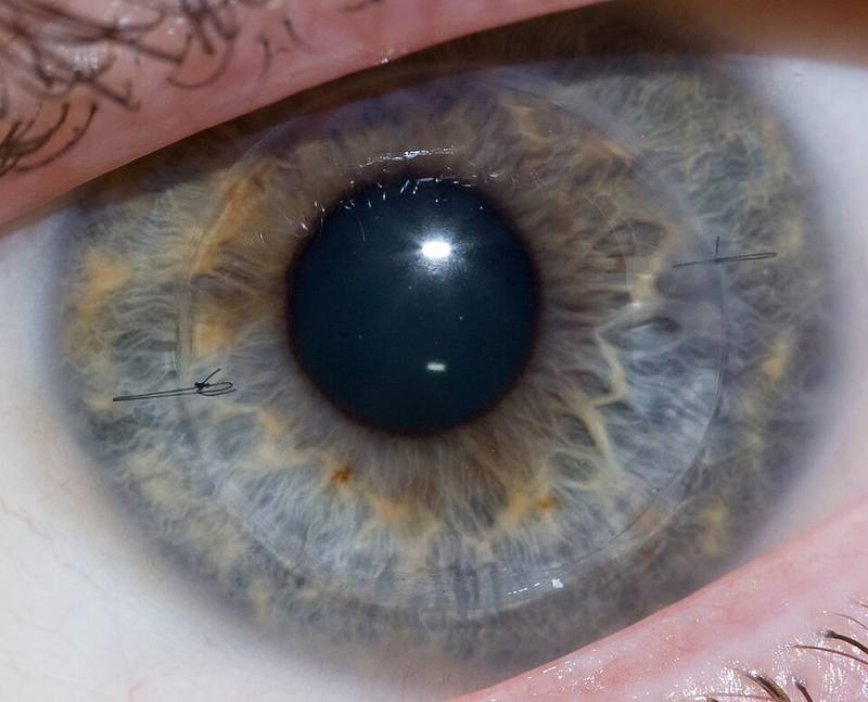
Corneal transplantation replaces diseased corneal tissue with donor tissue: full thickness in penetrating keratoplasty (PK), or selectively in lamellar techniques such as deep anterior lamellar keratoplasty (DALK) for stromal disease and endothelial keratoplasty (EK: DSAEK and DMEK) for endothelial disease [S1] [S2]. Lamellar techniques reduce rejection compared with PK [S2] [S10] [S12]. Graft rejection is the most common cause of late graft failure after PK [S4].
Key points - For Fuchs dystrophy and bullous keratopathy, EK appears to give higher 5-year graft survival and lower rejection than PK, on low-level evidence [S10]. - DMEK gives better visual acuity than ultrathin DSAEK but needs more re-bubbling [S9] [S11]. - For keratoconus, DALK and PK give comparable vision; DALK has less rejection [S2] [S12]. - After PK, pseudophakic or aphakic corneal oedema and prior glaucoma raise the risk of graft failure [S3]. - Evidence for immunosuppressants to prevent rejection after PK is largely low quality [S4].
Evidence last checked: 2026-10-17 (PubMed abstracts).
Red flags
- A graft patient with a red eye, reduced vision, light sensitivity or pain: possible rejection or infection; graft rejection is the most common cause of late failure after PK [S4] (see "Refer when").
- Raised eye pressure on post-graft steroids. After EK, ocular hypertension occurred in 20% with standard steroids and 7% with soft steroids [S7].
- Glaucoma history. 10-year graft failure after PK was 58% with prior glaucoma surgery and medication versus 22% without [S3].
- Persistent epithelial defect after keratoplasty [S2].
Recognise it: who needs a graft
- Fuchs endothelial dystrophy: endothelial loss leads to corneal oedema and reduced vision, and replacement with donor tissue is the only successful surgical treatment [S1].
- Pseudophakic or aphakic corneal oedema (bullous keratopathy) [S3] [S5].
- Keratoconus: about 10 to 15% of patients diagnosed with keratoconus require transplantation [S2].
Confirm it: choosing the technique
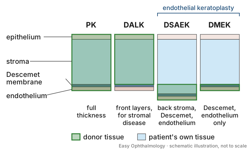
- Endothelial disease (EK vs PK). A Cochrane review of 3 small RCTs found no strong evidence of a difference in final vision between EK and PK, fewer higher-order aberrations but greater endothelial cell loss after EK [S1]. A systematic review of 50 cohort studies found 5-year graft survival of 0.93 to 0.99 after DMEK versus 0.69 to 0.98 after PK in Fuchs dystrophy, and 5-year rejection of 1.7 to 2.6% after DMEK, 5.0 to 7.9% after DSEK and 11.0 to 28.7% after PK [S10].
- DMEK vs ultrathin DSAEK. A meta-analysis of 7 studies found better 12-month vision after DMEK (SMD 0.50) but more re-bubbling (RR 0.33 favouring UT-DSAEK); other outcomes did not differ significantly [S9]. A review of 4 systematic reviews reached the same conclusion [S11].
- Keratoconus (DALK vs PK). A Cochrane review of 2 RCTs found no difference in vision and less rejection with DALK (OR 0.33, moderate certainty) [S2]. A 2025 meta-analysis of 47 studies (27 018 eyes) found comparable vision, more rejection with PK, but lower graft failure with PK than DALK [S12].
Manage it
Before acting on this section, check the national and local guidelines, product information and regulatory approvals that apply where you practise. See the medical disclaimer.
Tier A (primary care / no specialist)
- Refer any grafted eye that becomes red, painful or blurred (see "Refer when"); rejection is the most common cause of late graft failure after PK [S4].
- Post-graft drug regimens are not covered by these abstracts: follow the graft surgeon's plan and local formularies.
Tier B (general eye clinic)
- Check intraocular pressure in patients on post-graft steroids; soft steroids caused less ocular hypertension than standard steroids after EK [S7].
- Monitor endothelial cell density and corneal thickness where possible: after PK, lower density and higher thickness during follow-up were associated with later failure, although most corneas remained clear at 10 years [S3].
Tier C (specialist centre)
- Rejection prophylaxis after EK. In 49 studies (12 893 eyes), DMEK had less rejection than DS(A)EK; standard and soft steroids did not differ in preventing rejection after DMEK [S7].
- Immunosuppression after PK. A Cochrane review of 6 trials found low-quality evidence: systemic mycophenolate may not improve clear graft survival but may reduce rejection, and topical cyclosporine probably makes little difference to rejection after normal-risk grafts [S4].
- Donor and surgical factors. In the Cornea Preservation Time Study (1330 DSAEK eyes), 94% of grafts were clear at 3 years; diabetic donor tissue and operative complications increased early failure, and PACE increased late failure compared with Fuchs dystrophy [S5]. In the Cornea Donor Study, 10-year PK failure was 37% for PACE versus 20% for Fuchs dystrophy [S3].
- Surgeon training in low- and middle-income settings. In an Indian survey, DMEK was performed by 55% of responding surgeons, only 40% of whom had formal training; lack of training was the main barrier and DSEK remained the most common EK [S8]. For DALK, big-bubble technique was most preferred, and 50% of surgeons felt more than 20 cases were needed to pass the learning curve [S6]. These are surveys with low response rates.
Refer when
| Situation | Urgency |
|---|---|
| Grafted eye with redness, pain, photophobia or reduced vision (possible rejection or infection) | Same-day to the graft surgeon or cornea service [G1] |
| Raised intraocular pressure on post-graft steroids | Urgent to an eye clinic; timing per your local service or specialist advice [S7] |
| Corneal oedema or scarring reducing vision, suitable for grafting | Routine to a cornea service; timing per your local service or specialist advice |
Pitfalls
- Treating a red grafted eye as simple conjunctivitis [S4].
- Assuming DMEK is always best: it gives better vision but more re-bubbling [S9] [S11].
- Forgetting glaucoma and PACE as risk factors for failure [S3] [S5].
- Missing steroid-induced ocular hypertension [S7].
Evidence notes
| Source | Date | Design |
|---|---|---|
| [S1] | 2014 | Cochrane systematic review, 3 RCTs |
| [S2] | 2014 | Cochrane systematic review, 2 RCTs |
| [S3] | 2015 | Cornea Donor Study, multicentre masked trial, 1090 participants |
| [S4] | 2015 | Cochrane systematic review, 6 RCTs |
| [S5] | 2018 | Cohort within the Cornea Preservation Time RCT, 1330 eyes |
| [S6] | 2021 | Survey of Indian corneal surgeons (DALK) |
| [S7] | 2022 | Systematic review and meta-analysis, 49 studies |
| [S8] | 2022 | Survey of Indian corneal surgeons (DMEK) |
| [S9] | 2023 | Meta-analysis, 7 studies |
| [S10] | 2023 | Systematic review of 50 cohort studies |
| [S11] | 2024 | Review of 4 systematic reviews |
| [S12] | 2025 | Meta-analysis, 47 studies |
- Sources conflict on graft failure after DALK vs PK: no difference in the Cochrane review [S2], lower failure with PK in a larger observational meta-analysis [S12].
- The EK vs PK long-term data are from cohort studies with low level of evidence [S10]; the RCTs were small and lacked masking [S1].
- Most trials come from high-income countries; the two Indian sources are surveys [S6] [S8].
- Not covered by these sources: eye banking and donor supply in low-resource settings, suture management, treatment regimens for acute rejection, and keratoprosthesis.
Related articles
References
See the numbered source list below. Each marker [S#] links to its PubMed and DOI record.
Sources
- [S1] Nanavaty MA, Wang X, Shortt AJ Endothelial keratoplasty versus penetrating keratoplasty for Fuchs endothelial dystrophy. The Cochrane database of systematic reviews. (2014). PubMed 24526345 · doi:10.1002/14651858.cd008420.pub3
- [S2] Keane M, Coster D, Ziaei M, Williams K Deep anterior lamellar keratoplasty versus penetrating keratoplasty for treating keratoconus. The Cochrane database of systematic reviews. (2014). PubMed 25055058 · doi:10.1002/14651858.cd009700.pub2
- [S3] Writing Committee for the Cornea Donor Study Research Group, Sugar A, Gal RL, Kollman C, Raghinaru D, Dontchev M, et al. Factors associated with corneal graft survival in the cornea donor study. JAMA ophthalmology. (2015). PubMed 25322173 · doi:10.1001/jamaophthalmol.2014.3923
- [S4] Abudou M, Wu T, Evans JR, Chen X Immunosuppressants for the prophylaxis of corneal graft rejection after penetrating keratoplasty. The Cochrane database of systematic reviews. (2015). PubMed 26313245 · doi:10.1002/14651858.cd007603.pub2
- [S5] Terry MA, Aldave AJ, Szczotka-Flynn LB, Liang W, Ayala AR, Maguire MG, et al. Donor, Recipient, and Operative Factors Associated with Graft Success in the Cornea Preservation Time Study. Ophthalmology. (2018). PubMed 30098353 · doi:10.1016/j.ophtha.2018.08.002
- [S6] Fogla R, Sahay P, Sharma N Preferred practice pattern and observed outcome of deep anterior lamellar keratoplasty - A survey of Indian corneal surgeons. Indian journal of ophthalmology. (2021). PubMed 34011739 · doi:10.4103/ijo.ijo_3067_20
- [S7] Magnier F, Dutheil F, Pereira B, Watson SL, Baker JS, Chiambaretta F, et al. Preventive treatment of allograft rejection after endothelial keratoplasty: A systematic review and meta-analysis. Acta ophthalmologica. (2022). PubMed 35411680 · doi:10.1111/aos.15154
- [S8] Sahay P, Fogla R, Sharma N Preferred practice pattern for Descemet membrane endothelial keratoplasty surgeries: A survey of Indian corneal surgeons. Indian journal of ophthalmology. (2022). PubMed 35918952 · doi:10.4103/ijo.ijo_244_22
- [S9] Maier AB, Milek J, Joussen AM, Dietrich-Ntoukas T, Lichtner G Systematic Review and Meta-analysis: Outcomes After Descemet Membrane Endothelial Keratoplasty Versus Ultrathin Descemet Stripping Automated Endothelial Keratoplasty. American journal of ophthalmology. (2023). PubMed 36220351 · doi:10.1016/j.ajo.2022.09.013
- [S10] Lemaitre D, Tourabaly M, Borderie V, Dechartres A Long-term Outcomes After Lamellar Endothelial Keratoplasty Compared With Penetrating Keratoplasty for Corneal Endothelial Dysfunction: A Systematic Review. Cornea. (2023). PubMed 37185592 · doi:10.1097/ico.0000000000003240
- [S11] Moura-Coelho N, Papa-Vettorazzi R, Reyes A, Cunha JP, Güell JL Ultrathin DSAEK versus DMEK - Review of systematic reviews. European journal of ophthalmology. (2024). PubMed 37964555 · doi:10.1177/11206721231214605
- [S12] A Awad A, M Elettreby A, A Abo Elnaga A, A Alsaied M, M Aboelkhier M, Saleh MM, et al. Penetrating Keratoplasty versus Deep Anterior Lamellar Keratoplasty for Keratoconus: A Systematic Review and Meta-Analysis of 27,018 Eyes. Seminars in ophthalmology. (2025). PubMed 39943883 · doi:10.1080/08820538.2025.2457583
Guidelines
- [G1] NHS Scotland (Community Eyecare Guidelines, eyes.nhs.scot). 5.6 Transplant Rejection, Corneal (2026). https://www.eyes.nhs.scot/eyecare-guidelines/section-5-corneal/56-transplant-rejection-corneal/