Educational content only. Not personalised medical advice. Clinical decisions remain the responsibility of the treating clinician.
Diabetic retinopathy screening programmes
Diabetic retinopathy (DR) screening aims to detect retinopathy before it threatens sight so it can be treated in time [S2] [S4]. Retinal photography, graded by trained staff or artificial intelligence (AI), is accurate enough for programmes in low-resource settings [S3] [S10]. The weakest link is usually attendance: adherence was 39.3% in low- and middle-income countries [S7].
Key points - Non-mydriatic two-field photography, dilating only those with ungradable images, is a pragmatic way to start screening in low-income settings [S3] [S6]. - AI grading in developing countries had pooled sensitivity 0.933 and specificity 0.903 for referable DR [S10]. - Quality improvement interventions increased screening attendance by 12% over usual care (low-certainty evidence), but none of the 66 RCTs was from a low- or middle-income country [S1]. - Health education and knowledge about DR were linked to higher uptake; lack of skilled staff and imaging infrastructure were the main provider barriers [S4]. - Low-risk patients may be screened less often: intervals of 24 to 48 months for no or mild non-proliferative DR are supported by one national guideline [S2].
Evidence last checked: 2026-10-17 (PubMed abstracts).
Red flags: signs that need urgent action
- In a person: proliferative DR or diabetic macular oedema (DMO) on a screening image; globally, 6% of people with diabetes had proliferative DR and 11% sight-threatening DR [S8].
- In a person: ungradable images; dilate and re-image rather than report as normal [S3].
- In a programme: low attendance, especially in Africa (36.1%) [S7].
- In a programme: groups left behind; younger age, lower education, rural residence, lower income and no health insurance were linked to lower adherence [S7].
- In a programme: referrals with nowhere to go; lack of skilled human resources and imaging infrastructure are the main provider barriers [S4].
Recognise it: who to screen
- All people with diabetes. Pooled prevalence from retinal photography studies: any DR 23%, proliferative DR 6%, DMO 5%, sight-threatening DR 11% [S8].
- Children and adolescents with type 1 diabetes. Mean prevalence 9.5%, higher in low- and lower-middle-income countries; most cases occurred after five years of diabetes and after 11 years of age, and HbA1c ≥9% was linked to higher prevalence [S12]. Sight-threatening retinopathy was rare and largely confined to older adolescents with longer duration [S12].
- Those least likely to attend (see red flags) need targeted effort [S7].
Confirm it: the screening test
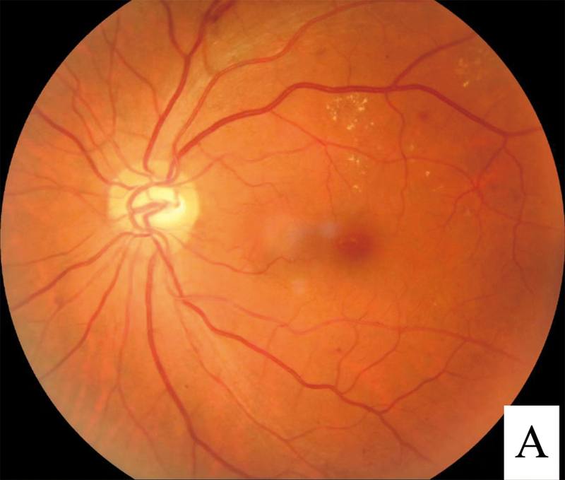
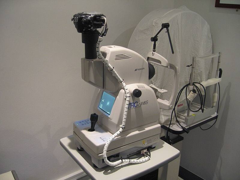
- Grading scale: the International Clinical Retinopathy Disease Severity Scale is recommended by the Danish guideline [S2].
- Fields: sensitivity and specificity rise with the number of photographic fields [S6]. For referable DR, sensitivity ranged from 76% with one field to 93% with wide-angle photography [S6].
- Pupil dilation: after excluding ungradable images there was no difference in sensitivity between non-mydriatic and mydriatic methods (86%) [S3]. In children, mydriatic screening detected more retinopathy than non-mydriatic (11.5% vs 4.4%) [S12]. The Danish guideline requires at minimum mydriatic two-field disc- and macula-centred images [S2]; these sources differ on mydriasis.
- Macular oedema: optical coherence tomography (OCT) can add diagnostic accuracy when clinically significant DMO is suspected [S2].
- Smartphone imaging: pooled sensitivity 87% and specificity 94% for any DR, 91% and 89% for referral-warranted DR, but only 39% sensitivity for mild NPDR [S5].
- AI grading: in prospective real-world studies, AUROC was 0.959 for referable DR and 0.974 for vision-threatening DR [S11]. In developing countries, AI pooled sensitivity was 0.891 and specificity 0.936 for vision-threatening DR, with a very wide confidence interval for sensitivity (0.393 to 0.990) [S10]. Camera type affected performance [S10].
Manage it: running a programme at each resource tier
Before acting on this section, check the national and local guidelines, product information and regulatory approvals that apply where you practise. See the medical disclaimer.
Tier A (primary care / no specialist)
- Offer screening at the diabetes clinic; nearby screening facilities enable uptake [S4].
- Trained non-ophthalmologists and general physicians using portable fundus cameras showed good agreement with reference standards [S10].
- Educate every person with diabetes about DR: health education was linked to higher uptake (OR 4.3) [S4].
- Use reminders and other quality improvement strategies; both screening-specific and general diabetes care interventions worked, especially where attendance was low [S1].
Tier B (general eye clinic)
- Grade images, or supervise AI output, and run the referral pathway [S10] [S11].
- Dilate and re-image when images are ungradable [S3].
- Consider individualised intervals for low-risk patients only if local guidance allows: 24 to 48 months for no or mild NPDR, and 12 to 24 months for moderate NPDR with HbA1c ≤53 mmol/mol and blood pressure ≤130/80 mmHg [S2].
Tier C (specialist centre and planners)
- Treat referable and sight-threatening DR; confirm DMO with OCT [S2].
- Plan staff, training and imaging infrastructure, the main provider-side barriers [S4].
- AI and telemedicine screening were cost-effective in seven studies from China, Thailand, Brazil and India [S9]; in middle-income countries results were mixed, depending on compliance, glycaemic control and initial costs [S11].
- Validate any screening method locally, since most diagnostic accuracy data come from high-income settings [S3].
Refer when
| Situation | Urgency |
|---|---|
| Proliferative DR, vitreous haemorrhage or sudden vision loss in a person with diabetes | Urgent to a retina or laser service; timing per your local service or specialist advice |
| Suspected DMO or severe NPDR on screening | Within a week to a few weeks, to an eye clinic with OCT [EIC expert opinion] |
| Ungradable images after dilation | Routine, for examination by an eye professional; timing per your local service or specialist advice [EIC expert opinion] |
| No or mild DR | Routine rescreening, interval as set by local guidance [S2] |
Pitfalls
- Reporting ungradable images as normal [S3].
- Relying on single-field images: sensitivity for referable DR can fall to 76% [S6].
- Trusting smartphone imaging to detect mild disease [S5].
- Deploying AI without checking camera type and image quality locally [S10] [S11].
- Building screening without staff and treatment capacity downstream [S4].
- Assuming high-income attendance evidence transfers unchanged [S1].
Evidence notes
| Source | Date | Design |
|---|---|---|
| [S1] | 2018 | Cochrane systematic review, 66 RCTs |
| [S2] | 2018 | National guideline based on systematic review (Denmark) |
| [S3] | 2018 | Diagnostic accuracy meta-analysis, 21 studies |
| [S4] | 2019 | Systematic review, 77 studies, mostly cross-sectional |
| [S5] | 2020 | Diagnostic accuracy meta-analysis, 9 studies, 1430 participants |
| [S6] | 2023 | Diagnostic accuracy meta-analysis, 62 studies |
| [S7] | 2024 | Systematic review and meta-analysis, 77 studies, 28 countries |
| [S8] | 2025 | Review and meta-analysis of prevalence, 42 studies |
| [S9] | 2025 | Systematic review of 7 economic evaluations |
| [S10] | 2025 | Systematic review and meta-analysis, 25 studies in developing countries |
| [S11] | 2026 | Systematic review and meta-analysis, 47 studies |
| [S12] | 2026 | Systematic review, 62 observational and interventional studies |
- Sources conflict on mydriasis: equal sensitivity without it in adults after excluding ungradable images [S3], better detection with it in children [S12], and the Danish guideline requires it [S2].
- The Danish guideline judged automated screening "not ready for implementation" in 2018 [S2]; later meta-analyses report high AI accuracy [S10] [S11].
- Diagnostic accuracy data come mostly from high-income settings [S3] [S4]; insights into low-income countries were limited [S11].
- No randomised trials compared screening strategies in children [S12].
- Global prevalence trends are hard to interpret because of differences in study design [S8].
- Not covered by these sources: treatment of DR and DMO, specific referral timeframes, and grader training standards.
Related articles
- Proliferative Diabetic Retinopathy: Laser, Anti-VEGF and Vitrectomy
- Artificial Intelligence in Retina and OCT
- Smartphone and portable fundus imaging
References
See the numbered source list below. Each marker [S#] links to its PubMed and DOI record.
Sources
- [S1] Lawrenson JG, Graham-Rowe E, Lorencatto F, Burr J, Bunce C, Francis JJ, et al. Interventions to increase attendance for diabetic retinopathy screening. The Cochrane database of systematic reviews. (2018). PubMed 29333660 · doi:10.1002/14651858.cd012054.pub2
- [S2] Grauslund J, Andersen N, Andresen J, Flesner P, Haamann P, Heegaard S, et al. Evidence-based Danish guidelines for screening of diabetic retinopathy. Acta ophthalmologica. (2018). PubMed 30311394 · doi:10.1111/aos.13936
- [S3] Piyasena MMPN, Murthy GVS, Yip JLY, Gilbert C, Peto T, Gordon I, et al. Systematic review and meta-analysis of diagnostic accuracy of detection of any level of diabetic retinopathy using digital retinal imaging. Systematic reviews. (2018). PubMed 30404665 · doi:10.1186/s13643-018-0846-y
- [S4] Piyasena MMPN, Murthy GVS, Yip JLY, Gilbert C, Zuurmond M, Peto T, et al. Systematic review on barriers and enablers for access to diabetic retinopathy screening services in different income settings. PloS one. (2019). PubMed 31013274 · doi:10.1371/journal.pone.0198979
- [S5] Tan CH, Kyaw BM, Smith H, Tan CS, Tudor Car L Use of Smartphones to Detect Diabetic Retinopathy: Scoping Review and Meta-Analysis of Diagnostic Test Accuracy Studies. Journal of medical Internet research. (2020). PubMed 32347810 · doi:10.2196/16658
- [S6] Yan W, McGuinness M, Chakrabarti R, Fotis K, Finger RP Comparison of Photographic Screening Methods for Diabetic Retinopathy - A Meta-analysis. Ophthalmic epidemiology. (2023). PubMed 35599625 · doi:10.1080/09286586.2022.2065311
- [S7] Rahmati M, Smith L, Boyer L, Fond G, Yon DK, Lee H, et al. Factors Affecting Global Adherence for the Uptake of Diabetic Retinopathy Screening: A Systematic Review and Meta-Analysis. American journal of ophthalmology. (2024). PubMed 39094991 · doi:10.1016/j.ajo.2024.07.028
- [S8] Owens DR, Gurudas S, Sivaprasad S, Zaidi F, Tapp R, Kazantzis D, et al. IDF diabetes Atlas: A worldwide review of studies utilizing retinal photography to screen for diabetic retinopathy from 2017 to 2024 inclusive. Diabetes research and clinical practice. (2025). PubMed 40578519 · doi:10.1016/j.diabres.2025.112346
- [S9] Andayani A, Hendrawan KA, Yudistira, Suryathi NMA, Ernawati T Economic Evaluation of Diabetic Retinopathy Screening in Developing Countries: A Systematic Review. Korean journal of ophthalmology : KJO. (2025). PubMed 40976613 · doi:10.3341/kjo.2025.0047
- [S10] Yudistira Y, Hendrawan KA, Andayani A, Suryathi NMA, Ernawati T, Gunawan ACV, et al. Diabetic Retinopathy Screening Approaches in Developing Countries: A Systematic Review and Meta-Analysis. Turkish journal of ophthalmology. (2025). PubMed 41142633 · doi:10.4274/tjo.galenos.2025.32916
- [S11] Ran AR, Ding JL, Tang Z, Lam C, Nguyen TX, Zhou J, et al. Real-World Prospective Validation and Economic Evaluation of Deep Learning- Based Diabetic Retinopathy Detection From Fundus Photographs: A Systematic Review and Meta-analysis. Diabetes care. (2026). PubMed 41259706 · doi:10.2337/dc25-1493
- [S12] Puli L, Piona C, Mancioppi V, Passanisi S, Kavanagh S, Donaghue KC Optimal screening practices for diabetic retinopathy in children and adolescents with type 1 diabetes: a systematic review to inform global screening guidance. Diabetes research and clinical practice. (2026). PubMed 42373008 · doi:10.1016/j.diabres.2026.113385