Educational content only. Not personalised medical advice. Clinical decisions remain the responsibility of the treating clinician.
Keratoconus and corneal cross-linking
Keratoconus is a lifelong, sight-threatening corneal ectasia that usually begins in the second to fourth decade of life and causes vision loss through thinning, irregular astigmatism and scarring [S4] [S2]. Corneal collagen cross-linking (CXL) with riboflavin and ultraviolet A light is the only treatment shown to slow progression [S4]. Progression is fastest in young patients and steep corneas, so early detection matters [S2].
Key points - Untreated keratoconus steepened by a mean of 0.7 D in Kmax over 12 months in a meta-analysis of 11 529 eyes; younger age and Kmax above 55 D predicted more progression [S2]. - Eye rubbing is consistently associated with keratoconus (pooled OR 6.46), although causality is unproven [S3]. - There is no unified definition of progression; Kmax change is the most common criterion [S10]. - Epithelium-off CXL is the reference technique; no alternative (transepithelial or accelerated) has been shown to be better at halting progression [S4] [S9]. - In children, pooled progression after epithelium-off CXL was about 10% [S6].
Evidence last checked: 2026-10-17 (PubMed abstracts).
Red flags
- A young patient (under 17 years) or Kmax steeper than 55 D: higher risk of progression; closer follow-up and a lower threshold for CXL are advised [S2].
- Vision that can no longer be fully corrected with glasses in a young person: consider keratoconus [S7].
- Refractive surgery candidates. Missed keratoconus before refractive surgery can lead to ectasia [S7].
- Pain, redness or an infiltrate in the days after CXL. Infectious keratitis after epithelium-off CXL is uncommon but presented at a mean of 4.8 days, mostly bacterial [S5].
- Corneal perforation or scarring can occur in keratoconus [S4].
Recognise it
- Frequency. Reported as about 1 in 2000 people [S1] or 86 in 100 000 [S2].
- Risk factors. Eye rubbing (pooled OR 6.46 from six case-control studies) [S3].
- Symptoms. Gradual increase in corneal curvature and decrease in visual acuity, with impact on quality of life [S1].
Confirm it
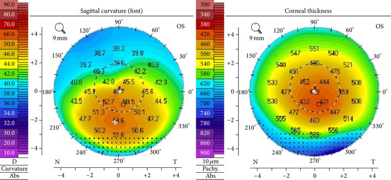
- Diagnosis is based on clinical examination and corneal imaging; early cases without clinical signs depend on topography or tomography interpreted by trained specialists [S7].
- AI tools. A Cochrane review of 63 studies found summary sensitivity 98.6% and specificity 98.3% for manifest keratoconus, and 90.0% and 95.5% for subclinical keratoconus, but the evidence was low to very low certainty [S7].
- Defining progression. Of 221 studies, 85.5% used Kmax, and most assessed progression over 6 to 12 months; criteria are not unified [S10].
Manage it
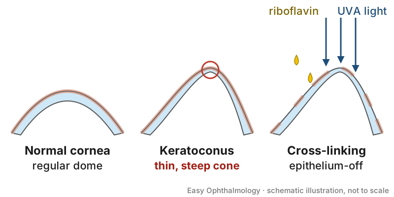
Before acting on this section, check the national and local guidelines, product information and regulatory approvals that apply where you practise. See the medical disclaimer.
Tier A (primary care / no specialist)
- Advise patients not to rub their eyes [S3].
- Refer young people whose vision is no longer fully corrected by glasses for corneal imaging [S7] (see "Refer when").
Tier B (general eye clinic)
- Refract, examine and arrange topography or tomography where available [S7].
- Monitor for progression, most commonly by Kmax change over 6 to 12 months [S10]; follow younger patients and steep corneas more closely [S2].
- Spectacles and contact lenses are not covered by these abstracts.
Tier C (specialist centre)
- CXL for progressive disease. A Cochrane review (2015) of 3 RCTs (219 eyes analysed) found treated eyes about 2 D less steep and with about 2 lines better uncorrected vision at 12 months, but evidence was very low quality [S1]. Adverse effects were not uncommon but mostly transient [S1].
- Epithelium-on versus epithelium-off. A Cochrane review of 13 RCTs found no clear difference in Kmax at 12 months or later (very low certainty); epithelium-off CXL caused slightly more haze or scarring (moderate certainty). It remains unknown whether any approach is better than epithelium-off [S4].
- Accelerated versus conventional. A meta-analysis of 14 RCTs found conventional CXL gave greater flattening and a deeper demarcation line, whereas accelerated CXL caused less thinning; visual outcomes did not differ [S9].
- Thin corneas (under 400 µm). In 29 mostly retrospective studies (470 eyes), modified CXL protocols flattened Kmax by 1.4 D at 12 months without reported major complications [S12].
- Children. In 35 studies (2186 eyes) most CXL protocols achieved stabilisation above 80% [S11]; pooled progression after epithelium-off CXL was 9.9% [S6].
- After CXL. Postoperative steroids and bandage contact lenses, and atopic or herpetic disease, were associated with infection; prophylactic antivirals are advised where appropriate [S5].
- Visual rehabilitation. A network meta-analysis suggests combinations of phakic intraocular lenses, intracorneal ring segments and CXL for regular corneas up to stage 3, and surface ablation with ring segments and CXL for irregular corneas [S8]. This is a complex synthesis and should be applied by specialists.
Refer when
| Situation | Urgency |
|---|---|
| Pain, redness or infiltrate after CXL | Same-day to the treating centre [S5] |
| Suspected or documented progression, especially under 17 years or Kmax above 55 D | Urgent to a cornea service; timing per your local service or specialist advice [S2] |
| Stable keratoconus with poor corrected vision | Routine to a cornea service; timing per your local service or specialist advice |
Pitfalls
- Waiting for progression in a child; younger patients progress faster [S2].
- Assuming transepithelial or accelerated CXL is equivalent in every respect [S4] [S9].
- Using a single Kmax reading as the only progression criterion [S10].
- Missing early keratoconus before refractive surgery [S7].
Evidence notes
| Source | Date | Design |
|---|---|---|
| [S1] | 2015 | Cochrane systematic review, 3 RCTs |
| [S2] | 2019 | Systematic review and meta-analysis, 11 529 eyes |
| [S3] | 2021 | Meta-analysis of 6 case-control studies |
| [S4] | 2021 | Cochrane systematic review, 13 RCTs |
| [S5] | 2021 | Systematic review of 26 publications |
| [S6] | 2022 | Systematic review and pooled analysis, 37 studies |
| [S7] | 2023 | Cochrane diagnostic accuracy review, 63 studies |
| [S8] | 2024 | Systematic review and network meta-analysis |
| [S9] | 2025 | Meta-analysis of 14 RCTs |
| [S10] | 2025 | Systematic review of 221 articles |
| [S11] | 2025 | Systematic review, 35 paediatric studies |
| [S12] | 2026 | Meta-analysis, mostly retrospective case series |
- The Cochrane CXL trials were at high risk of performance, detection and attrition bias and enrolled only adults [S1].
- The two prevalence estimates differ (1 in 2000 vs 86 in 100 000) [S1] [S2].
- Paediatric and thin-cornea data are largely observational with high heterogeneity [S6] [S12].
- Most AI studies were case-control designs at high risk of bias [S7].
- Not covered by these sources: spectacle and contact lens fitting, keratoplasty outcomes for keratoconus, cost and access to CXL in low-resource settings.
Related articles
- Corneal transplantation: DMEK, DSAEK and DALK
- Laser refractive surgery: LASIK, SMILE and PRK
- Contact lens complications and safe wear
References
See the numbered source list below. Each marker [S#] links to its PubMed and DOI record.
Sources
- [S1] Sykakis E, Karim R, Evans JR, Bunce C, Amissah-Arthur KN, Patwary S, et al. Corneal collagen cross-linking for treating keratoconus. The Cochrane database of systematic reviews. (2015). PubMed 25803325 · doi:10.1002/14651858.cd010621.pub2
- [S2] Ferdi AC, Nguyen V, Gore DM, Allan BD, Rozema JJ, Watson SL Keratoconus Natural Progression: A Systematic Review and Meta-analysis of 11 529 Eyes. Ophthalmology. (2019). PubMed 30858022 · doi:10.1016/j.ophtha.2019.02.029
- [S3] Sahebjada S, Al-Mahrouqi HH, Moshegov S, Panchatcharam SM, Chan E, Daniell M, et al. Eye rubbing in the aetiology of keratoconus: a systematic review and meta-analysis. Graefe's archive for clinical and experimental ophthalmology = Albrecht von Graefes Archiv fur klinische und experimentelle Ophthalmologie. (2021). PubMed 33484296 · doi:10.1007/s00417-021-05081-8
- [S4] Ng SM, Ren M, Lindsley KB, Hawkins BS, Kuo IC Transepithelial versus epithelium-off corneal crosslinking for progressive keratoconus. The Cochrane database of systematic reviews. (2021). PubMed 33765359 · doi:10.1002/14651858.cd013512.pub2
- [S5] Murchison CE, Petroll WM, Robertson DM Infectious keratitis after corneal crosslinking: systematic review. Journal of cataract and refractive surgery. (2021). PubMed 33769765 · doi:10.1097/j.jcrs.0000000000000620
- [S6] Achiron A, El-Hadad O, Leadbetter D, Hecht I, Hamiel U, Avadhanam V, et al. Progression of Pediatric Keratoconus After Corneal Cross-Linking: A Systematic Review and Pooled Analysis. Cornea. (2022). PubMed 34294638 · doi:10.1097/ico.0000000000002808
- [S7] Vandevenne MM, Favuzza E, Veta M, Lucenteforte E, Berendschot TT, Mencucci R, et al. Artificial intelligence for detecting keratoconus. The Cochrane database of systematic reviews. (2023). PubMed 37965960 · doi:10.1002/14651858.cd014911.pub2
- [S8] Niazi S, Doroodgar F, Hashemi Nazari S, Rahimi Y, Alió Del Barrio JL, Gatzioufas Z, et al. Refractive surgical approaches to keratoconus: A systematic review and network meta-analysis. Survey of ophthalmology. (2024). PubMed 38710236 · doi:10.1016/j.survophthal.2024.04.008
- [S9] Yeh CY, Chen KC, Chen YJ, Cheng SF Accelerated versus conventional corneal collagen cross-linking for keratoconus: A meta-analysis of randomized controlled trials. European journal of ophthalmology. (2025). PubMed 39539159 · doi:10.1177/11206721241298317
- [S10] Henriquez MA, Larco C, Izquierdo L Definition of Progressive Keratoconus: A Systematic Review. Cornea. (2025). PubMed 39671084 · doi:10.1097/ico.0000000000003777
- [S11] Morgado CR, Larivoir NB, Santos JF, Santhiago MR Corneal Cross-linking in Pediatric Populations: Systematic Review and Future Perspectives. Journal of refractive surgery (Thorofare, N.J. : 1995). (2025). PubMed 40923612 · doi:10.3928/1081597x-20250624-03
- [S12] Mohammadi F, McGuinness MB, Mustafa MZ, Chong EW, Daniell M Effectiveness and Safety of Cross-Linking in Keratoconus Patients With Corneal Thickness <400 µm: A Systematic Review and Meta-analysis. American journal of ophthalmology. (2026). PubMed 41016520 · doi:10.1016/j.ajo.2025.09.039